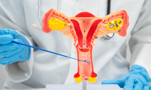
Understanding Vaginal Surgeries
Vaginal surgeries include reconstructive and aesthetic options tailored to individual needs. Common types involve repairing or tightening tissues affected by childbirth, aging, injury, or congenital conditions. These procedures fall under gynecology and increasingly under cosmetic gynecology, where the focus may include improving aesthetics alongside functionality.
1. Vaginal Hysterectomy
Vaginal hysterectomy involves removing the uterus through the vagina, avoiding abdominal incisions. It’s a minimally invasive procedure preferred for benign conditions like fibroids, abnormal bleeding, or prolapse. Performed under general or regional anesthesia, it typically lasts 1-2 hours. Surgeons access the uterus via the vaginal canal, ligate blood vessels, and detach supporting ligaments before extraction. Ovaries may be preserved or removed based on patient needs.
Surgical Technique
The surgery begins with the patient under general or regional anesthesia. The surgeon detaches the uterus from surrounding ligaments and blood vessels via the vagina, then removes it. Ovaries may be left intact or removed if necessary. The vaginal cuff is sutured closed to prevent complications. Post-operatively, patients are monitored for bleeding or infection, with emphasis on pelvic floor exercises to maintain support.
Recovery and Care
Patients typically stay in the hospital for one to two days. Activities like heavy lifting are restricted for several weeks to allow healing. Follow-up visits ensure proper recovery, focusing on hormonal balance if ovaries were preserved. This method is suitable for women with adequate vaginal access and no extensive adhesions.
2. Congenital Prolapse Surgeries
Congenital prolapse occurs due to inherent weaknesses in pelvic support structures, often linked to connective tissue disorders or anatomical anomalies present from birth. It may manifest as uterine, vaginal, or rectal descent, causing discomfort, urinary issues, or bowel problems. Surgical intervention aims to restore normal anatomy and function.
Surgical Approaches
Procedures vary based on the affected organ. For uterine prolapse, sacrohysteropexy attaches the uterus to the sacrum using mesh or sutures. Vaginal vault suspension techniques, like sacrospinous ligament fixation, provide support for post-hysterectomy cases. In rectal prolapse, rectopexy secures the rectum to the sacral bone. These surgeries often use minimally invasive methods to reduce tissue trauma.
Postoperative Management
Recovery involves pain management and gradual return to activities. Pelvic floor therapy strengthens muscles to prevent recurrence. Patients are advised on lifestyle modifications, such as avoiding straining during bowel movements. Regular check-ups monitor for any signs of mesh erosion or organ dysfunction, ensuring long-term pelvic stability.
3. Uterovaginal Prolapse (Acquired) Surgeries
Causes and Diagnosis
Acquired uterovaginal prolapse develops over time due to factors like multiple childbirths, menopause, or chronic straining, weakening pelvic ligaments and muscles. It leads to the uterus and vagina descending, causing bulging sensations, pressure, or urinary retention. Diagnosis involves physical exams and imaging to assess severity.
Corrective Procedures
Surgical options include vaginal mesh implants for support or native tissue repairs like anterior and posterior colporrhaphy to tighten vaginal walls. For complete prolapse, sacrocolpopexy attaches the vaginal apex to the sacrum via laparoscopy. Hysterectomy may be combined if the uterus is involved, followed by vault suspension to maintain position.
Rehabilitation Strategies
Post-surgery, patients focus on wound care and infection prevention. Hormone therapy might be recommended for menopausal women to improve tissue elasticity. Physical therapy targets core and pelvic muscles for sustained support. Lifestyle advice includes weight management and proper lifting techniques to minimize strain on repaired structures.
4. Urinary Incontinence Surgeries
Types and Mechanisms
Urinary incontinence surgeries address involuntary urine leakage, often from stress (coughing, sneezing) or urge types. Stress incontinence results from weakened urethral support, while urge involves overactive bladder muscles. Procedures aim to restore continence by enhancing sphincter function or bladder control.
Common Surgical Interventions
For stress incontinence, mid-urethral slings place a synthetic tape under the urethra for support during pressure increases. Bulking agents inject materials around the urethra to narrow it. For urge incontinence, sacral neuromodulation implants a device to regulate nerve signals to the bladder. Bladder neck suspension elevates the urethra via sutures.
Follow-Up and Lifestyle Integration
After surgery, bladder training exercises help regain control. Patients monitor for urinary tract infections and adjust fluid intake. Long-term care includes avoiding caffeine and maintaining a healthy weight to support surgical outcomes. Periodic evaluations assess continence and address any adjustments needed for optimal function.
5. Old Complete Perineal Tear Repair
Nature of the Injury
An old complete perineal tear refers to a longstanding injury from childbirth, involving the perineum, anal sphincter, and rectal mucosa, leading to fecal incontinence or pain. If untreated initially, it may cause chronic issues like gas leakage or difficulty with bowel control, necessitating delayed surgical correction.
Repair Techniques
Surgery involves dissecting scarred tissue and reconstructing layers. The anal sphincter is overlapped and sutured for strength, while the rectal lining is repaired to restore integrity. Perineorrhaphy tightens the perineal body. End-to-end anastomosis may be used for sphincter reconnection, often under anesthesia with bowel preparation beforehand.
Healing and Supportive Measures
Postoperative care emphasizes stool softeners to prevent straining and wound hygiene to avoid infection. Biofeedback therapy trains sphincter muscles for better control. Patients gradually resume activities, with dietary fiber increases for regular bowel habits. Ongoing assessments ensure functional improvement and address any residual symptoms.
6. Vaginoplasty (Medical Indications Only)
Medical Necessities
Vaginoplasty for medical reasons reconstructs or creates a vagina in cases of congenital absence (like Mayer-Rokitansky-Küster-Hauser syndrome), post-trauma, or after cancer treatments removing vaginal tissue. It restores functionality for urination, menstruation, or intercourse, focusing on anatomical and physiological needs rather than aesthetics.
Operative Methods
Techniques include using skin grafts from the thigh or peritoneal lining to form the vaginal canal. The McIndoe procedure involves placing a mold covered in graft material into a created space, allowing epithelialization. Intestinal vaginoplasty uses a segment of bowel for a self-lubricating canal in complex cases. Surgery requires precise dissection to avoid nerve damage.
Recovery Protocols
Dilators are used postoperatively to maintain depth and width, with regular application for months. Hormone replacement may support tissue health in certain conditions. Psychological support addresses body image adjustments. Follow-ups monitor for stenosis or fistulas, ensuring the reconstructed vagina functions effectively in daily life.
7. Open Surgeries
Indications for Open Approach
Open surgeries in gynecology involve larger abdominal incisions for direct access, used when minimally invasive methods are unsuitable, such as in extensive adhesions, large tumors, or emergencies. Common for ovarian cystectomies, myomectomies, or staging in cancers, providing tactile feedback and comprehensive visualization.
Procedural Steps
Under general anesthesia, a midline or transverse incision exposes the pelvis. The surgeon manipulates organs manually, excising pathology like fibroids or cysts while preserving fertility if desired. Hemostasis is achieved with sutures or clamps, and layers are closed meticulously to prevent hernias. Drains may be placed for fluid management.
Postoperative Considerations
Hospital stays extend to several days for monitoring vital signs and bowel function. Pain control uses medications, with early ambulation to reduce clot risks. Scar care and infection prevention are key. Rehabilitation includes gradual exercise, with nutritional support for healing. Long-term, patients watch for adhesions through symptom awareness.